Anatomy Of Ribs And Sternum / The thoracic cage - the ribs and sternum | Human Anatomy and Physiology Lab (BSB 141). This guide gives a general overview of the anatomy of the thoracic spine. Where is the sternum located? It lies on the anterior thoracic wall in the middle. Anterior view of the thoracic cage image source. Attach the ribs to the costal cartilages.
The sternum is a flat, long bone that forms the medial and anterior part of the thoracic cage. Wondering what the sternum is? They are ribbon like, elastic bony arches and flat in shape. They increase in length, curvature and amount of cartilage craniocaudally. Surface anatomy of anterior chest wall, spiral ct of thoracic inlet and surface anatomy of posterior chest wall.
Sternum and Ribs - HUMAN ANATOMY WEB SITE from mesa-anatomy.weebly.com Lessons on the bone markings of the ribs and sternum. Important clinical anatomy of the head, neck, and back. This often has little impact on function or treatment following injury but can however, cartilaginous connectors between the sternum and each of the upper six ribs assist with minor motions that occur with each breath. Learn all about this bone using our interactive anatomy image and detailed descriptions of its parts and function! Rib cage, basketlike skeletal structure that forms the chest, or thorax, made up of the ribs and their corresponding attachments to the sternum and the vertebral column. According to their attachment to the sternum, the ribs are classified. The sternum, commonly known as the breastbone, is a long, narrow flat bone that serves as the keystone of the rib cage and stabilizes the thoracic skeleton. The sternum or breastbone is a long flat bone located in the central part of the chest.
Sternum, costal cartilages, and ribs image source:
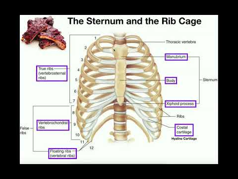
It consists of the ribs, the sternum, and the thoracic vertebrae, to which the ribs articulate. Describe the motion of the clavicle during upper limb movement and the sternum during respiration. This guide gives a general overview of the anatomy of the thoracic spine. There is a printable worksheet available for download here so you can take the quiz with pen and paper. Learn all about this bone using our interactive anatomy image and detailed descriptions of its parts and function! Important clinical anatomy of the head, neck, and back. Regional vertebrae (cervical, thoracic, lumbar), rib, sternum, os coxae, clavicle, scapula, humerus, ulna and radius for dr. The chest wall is formed from the sternum anteriorly, 12 pairs of ribs, costal cartilages and intercostal muscles laterally, and the thoracic vertebrae posteriorly. Try to be as accurate as you can with them. According to their attachment to the sternum, the ribs are classified. Interactive tutorials about the ribs and sternum bones, with labeled images and diagrams featuring the beautiful illustrations of getbodysmart. Attach the ribs to the costal cartilages. Ribs eight to ten are the false ribs and are connected to the sternum indirectly via the cartilage of the rib above them.
Learn more about the skeletal system with quizzes and labelling exercises. They are ribbon like, elastic bony arches and flat in shape. Interactive tutorials about the ribs and sternum bones, with labeled images and diagrams featuring the beautiful illustrations of getbodysmart. It has clear front, side, and back planes. The first portion, the manubrium, articulates with both the clavicle and first rib and is therefore.
Figure 1 from The anatomy of the ribs and the sternum and their relationship to chest wall ... from d3i71xaburhd42.cloudfront.net The rib cage is the arrangement of ribs attached to the vertebral column and sternum in the thorax of most vertebrates, that encloses and protects the vital organs such as the heart, lungs and great vessels. Join the sternum and clavicles. It connects to the ribs via cartilage and forms the front of the rib cage, thus helping to protect the heart, lungs, and major blood vessels from injury. Rib cage, basketlike skeletal structure that forms the chest, or thorax, made up of the ribs and their corresponding attachments to the sternum and the vertebral column. The final two pairs of ribs are floating ribs and the cartilage of these ribs tends to end within the abdominal musculature. Regional vertebrae (cervical, thoracic, lumbar), rib, sternum, os coxae, clavicle, scapula, humerus, ulna and radius for dr. The thoracic cage (rib cage) is the skeletal framework of the thoracic wall, which encloses the thoracic cavity. This guide gives a general overview of the anatomy of the thoracic spine.
We examined the thoracic vertebrae last lab, so here we will only examine the ribs and sternum.
Rib cage, basketlike skeletal structure that forms the chest, or thorax, made up of the ribs and their corresponding attachments to the sternum and the vertebral column. Sternum, costal cartilages, and ribs image source: The thoracic cage consists of the 12 thoracic vertebrae, the associated intervertebral discs, 12 pairs of ribs with their costal cartilages, and the sternum. This guide gives a general overview of the anatomy of the thoracic spine. Surface anatomy and surface markings bibliographic record list of illustrations subject index. Interactive tutorials about the ribs and sternum bones, with labeled images and diagrams featuring the beautiful illustrations of getbodysmart. The classification of human ribs. Costae are arranged in pairs and articulate with two successive vertebrae. Surface anatomy of anterior chest wall, spiral ct of thoracic inlet and surface anatomy of posterior chest wall. Lessons on the bone markings of the ribs and sternum. It discusses the specific anatomy of the ribs and costal cartilages, along with the sternum. Construct a robo skelly rib cage and the pelvis using the bucket method. The first portion, the manubrium, articulates with both the clavicle and first rib and is therefore.
The chest wall is formed from the sternum anteriorly, 12 pairs of ribs, costal cartilages and intercostal muscles laterally, and the thoracic vertebrae posteriorly. Learn about sternum thorax ribs anatomy with free interactive flashcards. An exception to this rule is that the first rib articulates with the first thoracic vertebra only. Each pair articulates with a. It connects to the ribs via cartilage and forms the front of the rib cage, thus helping to protect the heart, lungs, and major blood vessels from injury.
Human Organs Under Ribcage - Mocksure from i.ytimg.com Attach the ribs to the costal cartilages. Join the sternum and clavicles. The front plane is composed of the sternum and. The final two pairs of ribs are floating ribs and the cartilage of these ribs tends to end within the abdominal musculature. Try to be as accurate as you can with them. Lessons on the bone markings of the ribs and sternum. Sternum is a part of the skeletal system. Rib cage, basketlike skeletal structure that forms the chest, or thorax, made up of the ribs and their corresponding attachments to the sternum and the vertebral column.
Learn all about this bone using our interactive anatomy image and detailed descriptions of its parts and function!
They are ribbon like, elastic bony arches and flat in shape. Lessons on the bone markings of the ribs and sternum. Describe the bony and cartilaginous articulations of the sternum and clavicle. Surface anatomy and surface markings bibliographic record list of illustrations subject index. The sternum is a flat, long bone that forms the medial and anterior part of the thoracic cage. The rib cage surrounds the lungs and the heart, serving as an important means of bony protection for these vital organs. Individual ribs have a bony dorsal part, a body of rib, and ventral costal cartilage. Join the sternum and clavicles. An exception to this rule is that the first rib articulates with the first thoracic vertebra only. It lies on the anterior thoracic wall in the middle. Learn all about this bone using our interactive anatomy image and detailed descriptions of its parts and function! The sternum, commonly known as the breastbone, is a long, narrow flat bone that serves as the keystone of the rib cage and stabilizes the thoracic skeleton. The number is the same in both males and females.
Learn all about this bone using our interactive anatomy image and detailed descriptions of its parts and function! anatomy of ribs. This is an online quiz called ribs and sternum anatomy.
Share :
Post a Comment
for "Anatomy Of Ribs And Sternum / The thoracic cage - the ribs and sternum | Human Anatomy and Physiology Lab (BSB 141)"


)
Post a Comment for "Anatomy Of Ribs And Sternum / The thoracic cage - the ribs and sternum | Human Anatomy and Physiology Lab (BSB 141)"